Occasionally an arthroscopic debridement is needed if it does remain sore after 6 months.

COLLES FRACTURE PAIN FREE
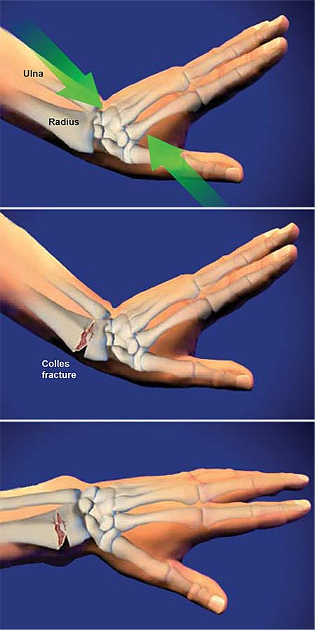
It is often torn at the same time as the distal radius fractures, but will almost always become pain free over time. There is a shock absorber tissue called the triangular fibrocartilage in this area. This is due to a soft tissue injury associated with the fracture. Pain at 10 days on a 11-point visual analog scale was significantly lower in the piroxicam group (2.1) compared with placebo (3.1, p<0.05 (actual p value not reported)). Ulnar-sided wrist pain after distal radius fracture can be the result of a variety of conditions, including DRUJ arthritis, DRUJ instability, and ulnocarpal abutment, all of which are conditions that may be caused or compounded by a malunion of the distal radius. Adolphson et al 17 randomized postmenopausal patients to piroxicam or placebo after suffering from a Colles’ fracture. Some pain on the little finger or ulnar side of the wrist is common for up to 6 months. A Colles’ fracture is the most common type. The small piece of bone is not floating around.

It often is healed with soft tissue rather than bone bridging the gap, but this rarely causes problems. This rarely needs any surgery or treatment. It is common to see a small pice of bone detached from the end of the ulna, the smaller little finger side forearm bone. Soft tissue injuries such as scapholunate ligament tears, and distal radioulnar joint instability need to be examined for. Complex FracturesĪt times there can be complex fracture involving the joint surface that need advanced techniques such as arthroscopic assistance to achieve a good result. In addition to RICE therapy you can use over-the-counter pain. Good function will usually result from a fracture healing in a good position, and with higher demands the fracture will need to be as close to original shape. You can manage the pain of a Colles fracture with RICE therapy (rest, ice, compress, elevate). Commonly if a reduction is needed, the fracture will have a plate and screws inserted to reliably hold it in place while it heals. Patients with a distal radius fracture typically present following an episode of trauma, complaining of immediate pain +/- deformity and sudden swelling around the fracture site. The number of cast changes was reported by the. Pain and stiffness were reported at the outpatient clinic at 3 months, and both pain and stiffness were found to be less in the FC group (Table 4).

30 Other studies have found that an increase in the ulnar variance was. Other symptoms can include: Bone that protrudes through your skin. Their wrist may even be noticeably out of alignment or deformed looking. Some fractures need an operation to reduce, or put the back in correct place, the fracture. Operative treatment due to loss of reduction of fracture was performed for four patients in the FC group and for seven patients in the VFUDC group. One study of 109 Colles’ fractures treated with closed reduction and casting determined that the most important factor for predicting ulnar wrist pain was incongruity of the distal radioulnar joint as a result of residual dorsal angulation of the radius. When a person has a Colles’ fracture they will have swelling and intense pain.


 0 kommentar(er)
